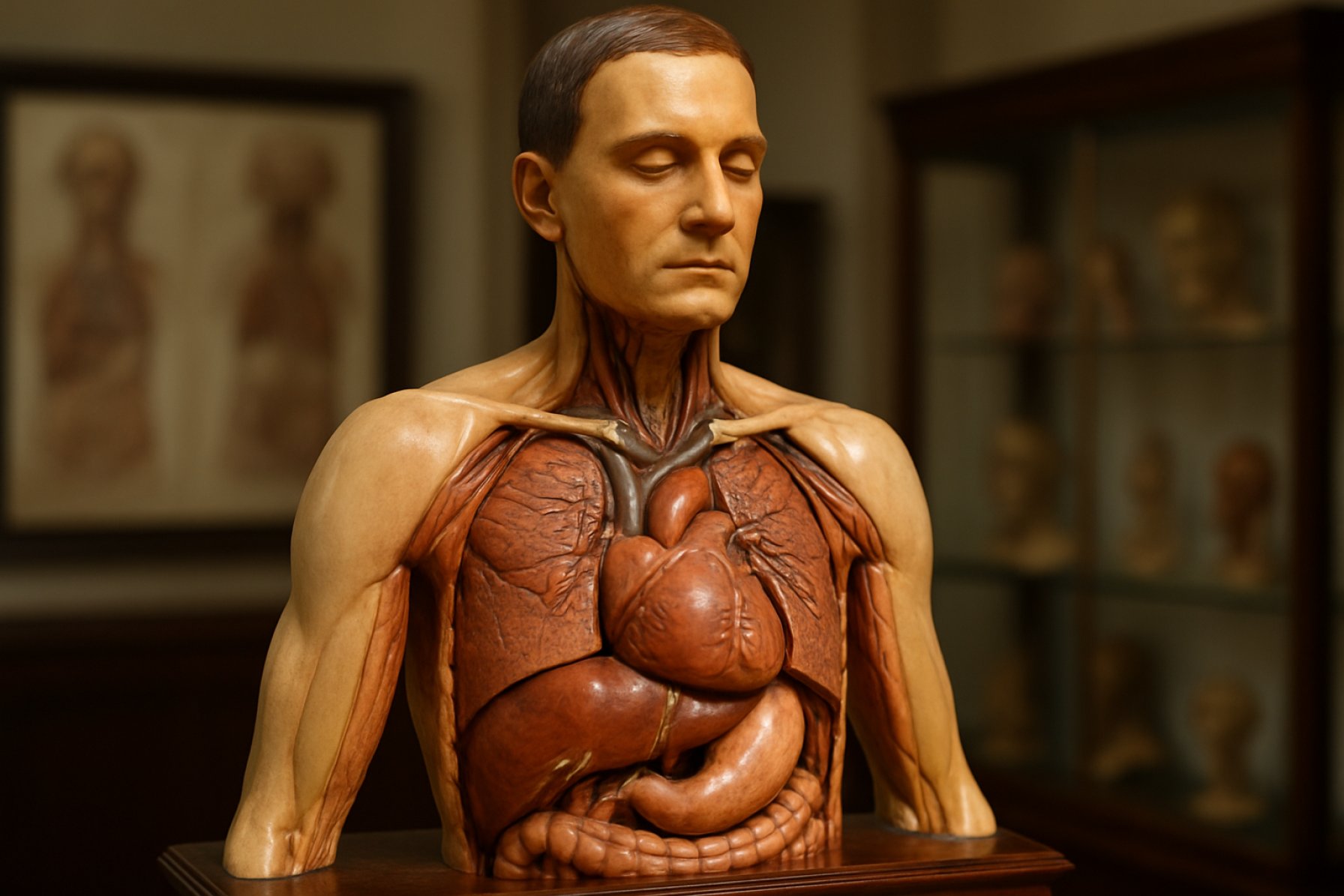
Wax Anatomical Models: Where Artistry Meets Medical Discovery. Explore the Intricate Craft and Enduring Legacy of These Lifelike Educational Tools.
- Origins and Historical Significance of Wax Anatomical Models
- Techniques and Materials: The Craft Behind the Models
- Educational Impact: Transforming Medical Training
- Artistic Value and Aesthetic Appeal
- Preservation and Conservation Challenges
- Notable Collections and Museums Worldwide
- Modern Relevance and Influence on Contemporary Medical Visualization
- Sources & References
Origins and Historical Significance of Wax Anatomical Models
The origins of wax anatomical models can be traced back to the late Renaissance and early modern period, particularly flourishing in Italy during the 17th and 18th centuries. These models emerged as a response to the growing demand for anatomical education and the limitations of cadaver dissection, which was often restricted by religious, legal, and practical constraints. Early centers of production included Florence and Bologna, where skilled artisans collaborated with anatomists to create lifelike representations of the human body. The Museo di Storia Naturale "La Specola" in Florence remains one of the most significant repositories of these historical models.
Wax anatomical models played a crucial role in the advancement of medical science and education. They allowed for the detailed study of human anatomy without the ethical and logistical challenges of using real bodies. These models were not only used for teaching medical students but also served as public exhibits, helping to demystify the human body for a broader audience. Their realism and durability made them invaluable tools for repeated study, and their creation required a unique blend of scientific knowledge and artistic skill. The University of Bologna and other European institutions became renowned for their collections, which contributed to the spread of anatomical knowledge across the continent.
Beyond their educational function, wax anatomical models are significant as cultural artifacts, reflecting the intersection of art, science, and society in early modern Europe. They illustrate the period’s fascination with the human body and the desire to render its mysteries visible and comprehensible, marking a pivotal moment in the history of medical visualization.
Techniques and Materials: The Craft Behind the Models
The creation of wax anatomical models, a practice that flourished from the Renaissance through the 19th century, required a sophisticated blend of artistry and scientific precision. Artisans and anatomists collaborated closely, employing a variety of techniques to achieve lifelike representations of human anatomy. The process typically began with detailed anatomical drawings or direct observation of dissections, ensuring accuracy in proportion and structure. Skilled modelers would then sculpt initial forms in clay or plaster, which served as the basis for negative molds.
The primary material, beeswax, was chosen for its malleability, translucency, and ability to capture fine detail. To enhance durability and achieve the desired coloration, wax was often mixed with resins, pigments, and sometimes animal fats. Layering techniques allowed for the simulation of skin, muscle, and internal organs, with each layer tinted to mimic the hues of living tissue. Glass eyes, human hair, and silk threads were sometimes incorporated to increase realism. The final models were often mounted on wooden bases and protected by glass cases.
Temperature control was crucial throughout the process, as wax becomes brittle when cold and overly soft when warm. Artisans used heated tools to shape and smooth surfaces, and fine brushes to apply color. The meticulous craftsmanship required for these models is exemplified by the collections at institutions such as the Museo di Storia Naturale "La Specola" and the Berlin Museum of Medical History at the Charité, where many historic wax models are still preserved and displayed.
Educational Impact: Transforming Medical Training
Wax anatomical models revolutionized medical education from the 17th through the 19th centuries, providing an invaluable alternative to human dissection at a time when cadaver access was limited and often controversial. These meticulously crafted models enabled students to study human anatomy in unprecedented detail, offering a three-dimensional, tactile resource that surpassed the limitations of two-dimensional illustrations. The lifelike coloration and textural accuracy of wax models allowed for the visualization of complex structures such as muscles, nerves, and organs, fostering a deeper understanding of spatial relationships within the body.
Institutions such as the Museo di Storia Naturale "La Specola" in Florence and the Gordon Museum of Pathology in London amassed extensive collections of these models, which became central to medical curricula. Wax models democratized anatomical knowledge, making it accessible to a broader audience, including women and laypeople who were often excluded from dissection rooms. Their durability and reusability also meant that rare pathological specimens could be studied repeatedly without ethical or practical concerns.
The educational impact of wax anatomical models extended beyond medical schools. They were used in public lectures and exhibitions, contributing to the popularization of anatomical science and the demystification of the human body. While modern technology has introduced digital and virtual alternatives, the historical role of wax models in transforming medical training remains a testament to their enduring educational value Public Health England.
Artistic Value and Aesthetic Appeal
Wax anatomical models, while primarily created for scientific and educational purposes, possess a remarkable artistic value and aesthetic appeal that has captivated viewers for centuries. The process of crafting these models required not only anatomical precision but also a high degree of artistic skill. Artisans meticulously sculpted wax to replicate the textures, colors, and forms of human tissues, organs, and even pathological conditions, often achieving a level of realism that rivaled fine art. The use of translucent wax allowed for lifelike representations of skin and internal structures, enhancing both the visual impact and educational utility of the models.
Many wax anatomical models were produced in the 18th and 19th centuries, particularly in centers such as Florence and Vienna, where artists collaborated closely with anatomists. The resulting works, such as those found in the Museo di Storia Naturale "La Specola", are celebrated not only for their scientific accuracy but also for their beauty and craftsmanship. The models often feature dramatic poses, expressive faces, and elaborate settings, blurring the line between scientific object and artistic masterpiece.
Today, these models are appreciated as cultural artifacts that reflect the intersection of art and science. Their aesthetic qualities—attention to detail, color, and form—continue to inspire contemporary artists and historians alike. Exhibitions in institutions such as the The Hunterian highlight the enduring fascination with the artistry embedded in these anatomical representations, underscoring their dual legacy as both educational tools and works of art.
Preservation and Conservation Challenges
Wax anatomical models, prized for their historical and educational value, present unique preservation and conservation challenges. The primary vulnerability of these models lies in the inherent properties of wax: it is highly sensitive to temperature fluctuations, light, and humidity. Even minor increases in ambient temperature can cause deformation, softening, or even melting, while low temperatures may render the wax brittle and prone to cracking. Relative humidity must be carefully controlled, as excessive moisture can promote mold growth and cause the wax to become sticky, whereas overly dry conditions may lead to desiccation and surface cracking (ICCROM).
Light exposure, particularly ultraviolet (UV) radiation, accelerates the degradation of both the wax and any pigments or surface finishes applied to the models. This can result in discoloration, fading, and loss of detail. Additionally, wax is susceptible to dust accumulation and insect infestation, especially if organic materials such as hair, textiles, or wood are incorporated into the models. Handling poses another risk, as fingerprints and pressure can leave permanent marks or cause structural damage (The British Museum).
Conservation efforts require a multidisciplinary approach, combining preventive measures—such as climate-controlled display cases, low-light environments, and minimal handling—with specialized restoration techniques. These may include consolidation of fragile areas, cleaning with non-invasive methods, and, in some cases, the use of reversible adhesives or fills. The complexity of these interventions underscores the importance of ongoing research and collaboration among conservators, scientists, and curators to ensure the long-term survival of these irreplaceable artifacts (The Getty Conservation Institute).
Notable Collections and Museums Worldwide
Wax anatomical models, renowned for their historical and educational significance, are preserved in several notable collections and museums worldwide. Among the most famous is the Museo di Storia Naturale "La Specola" in Florence, Italy, which houses the celebrated 18th-century collection of Clemente Susini. This museum features hundreds of life-sized and sectional wax models, meticulously crafted to illustrate human anatomy in extraordinary detail. Another significant institution is the Berlin Museum of Medical History at the Charité, which preserves a diverse array of anatomical waxworks, including pathological specimens and teaching models from the 19th and early 20th centuries.
In France, the Musée des Moulages de l’Hôpital Saint-Louis in Paris is renowned for its extensive collection of dermatological wax models, used historically for medical training and diagnosis. The United Kingdom’s Surgeons' Hall Museums in Edinburgh also display a significant number of wax anatomical models, reflecting the evolution of medical education in Britain.
Outside Europe, the Museum of Health Care at Kingston in Canada and the National Library of Medicine in the United States both maintain smaller but important collections, highlighting the global reach and enduring value of these artifacts. These museums not only preserve the artistry and scientific accuracy of wax anatomical models but also provide insight into the history of medical teaching and the visualization of the human body.
Modern Relevance and Influence on Contemporary Medical Visualization
Wax anatomical models, once essential tools for medical education in the pre-photographic era, continue to exert a significant influence on contemporary medical visualization. While digital technologies such as 3D modeling, virtual reality, and advanced imaging have largely supplanted traditional wax models in teaching, the principles underlying their creation—accuracy, tangibility, and didactic clarity—remain foundational in modern anatomical representation. The meticulous craftsmanship and lifelike detail of historical wax models set a benchmark for realism that contemporary digital models strive to emulate. Museums and medical schools still use these artifacts to inspire new generations of medical illustrators and educators, emphasizing the importance of tactile and visual learning experiences.
Moreover, the resurgence of interest in physical models for simulation-based learning has led to the development of synthetic and 3D-printed anatomical replicas, which draw directly from the legacy of wax modeling. These modern tools are used for surgical training, patient education, and interdisciplinary research, reflecting the enduring value of three-dimensional, hands-on learning. The preservation and digitization of historical wax models also contribute to current research, providing reference material for comparative anatomy and the history of medicine. Institutions such as the Science Museum Group and the Museo di Storia Naturale La Specola actively curate and display these models, highlighting their ongoing relevance. In sum, wax anatomical models continue to shape the evolution of medical visualization, bridging the gap between traditional craftsmanship and cutting-edge technology.
Sources & References
- University of Bologna
- Berlin Museum of Medical History at the Charité
- Gordon Museum of Pathology
- ICCROM
- The Getty Conservation Institute
- Surgeons' Hall Museums
- Museum of Health Care at Kingston
- National Library of Medicine
- Science Museum Group
